
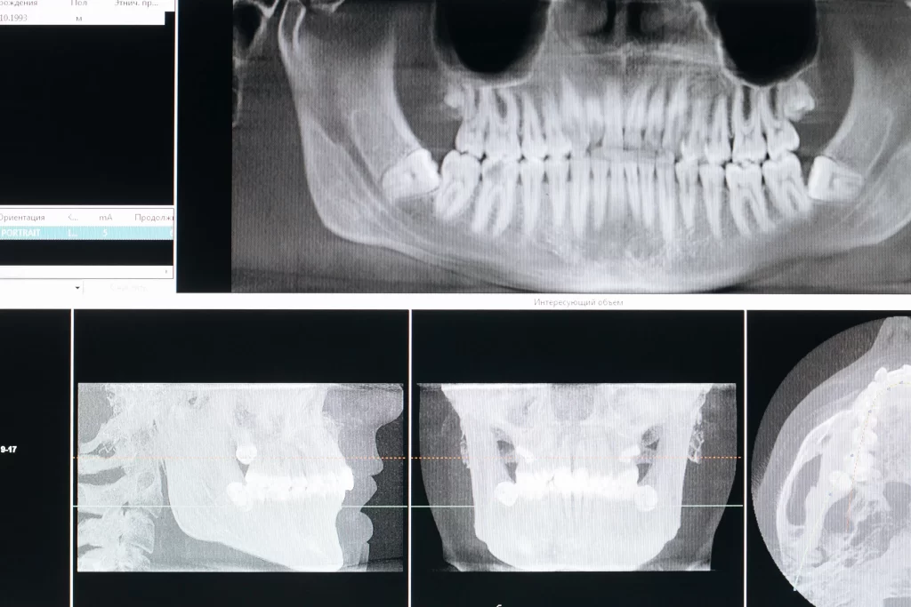
An OPG X-ray, short for Orthopantomogram, is a panoramic dental X-ray that creates a broad 2D view of the teeth, upper jaw, lower jaw and surrounding structures in a single image. Dentists use an OPG scan for overall dental assessment and treatment planning, including evaluating wisdom teeth, impacted or unerupted teeth, tooth development, jawbone changes and other visible abnormalities. It can also help with planning treatments such as braces, dental implants and oral surgery.
During an OPG, you stand or sit at the imaging machine while your head and teeth are positioned correctly. A rotating arm moves around your head to capture the image. The scan itself is brief and non-invasive, and it should not be painful.
Because an OPG uses X-rays, it involves ionising radiation. Dental X-rays should therefore be recommended when they are clinically appropriate, with exposure kept as low as reasonably achievable. An OPG also has an important limitation: it provides a broad 2D panoramic view, not unlimited detail. A dentist may recommend a focused intraoral X-ray such as an IOPA or a 3D scan such as CBCT when more detailed information is needed.
What Is an OPG X-Ray?
What does OPG stand for?
OPG stands for Orthopantomogram. It is also called a panoramic dental X-ray or panoramic radiograph.
Unlike an intraoral X-ray, where the detector is positioned inside the mouth to focus on a smaller area, an OPG captures a much wider view. The resulting image provides an overview of both jaws and the dentition, making it useful when a dentist needs to understand the bigger picture before deciding whether further imaging or treatment is needed.
What does an OPG X-ray show?
Depending on the image quality and the patient’s anatomy, an OPG can show:
OPG captures | Why it matters |
Upper and lower teeth | Provides an overall dental assessment |
Jawbones | Gives a broad view of bone structures |
Wisdom teeth | Helps assess position and possible impaction |
Developing or unerupted teeth | Helps assess tooth development and eruption |
Tooth roots | Provides general visibility of root position |
Surrounding jaw structures | Adds broader diagnostic context |
An OPG may also provide information about certain jaw abnormalities, impacted teeth, periodontal bone changes and other visible findings. However, the image should always be interpreted alongside the clinical examination rather than viewed in isolation.
What Is an OPG Scan Used For?
An OPG scan is mainly used when a dentist needs a broad view of the teeth and jaws. It can be useful for both diagnosis and treatment planning.
Common reasons dentists recommend an OPG
An OPG may be recommended to assess:
- Impacted or unerupted teeth
- Wisdom teeth and their position
- Tooth development, particularly when teeth are still developing or erupting
- Broad jawbone changes
- Suspected infection or other abnormalities
- Periodontal or supporting-bone changes
- Certain jaw conditions or visible abnormalities
- The overall position of teeth before orthodontic treatment
- The jaws and teeth before selected surgical procedures
An OPG can therefore be useful even when you do not have significant pain. Some dental problems, particularly impacted or developing teeth, may not cause obvious symptoms at the time they are assessed.
OPG for braces, implants and oral surgery
An OPG can contribute to treatment planning, although it is not automatically sufficient for every procedure.
Treatment or concern | Why an OPG may help |
Braces | Provides a broad view of tooth position and development |
Wisdom teeth | Shows the general position of impacted teeth |
Dental implants | Provides an initial overview before further imaging when required |
Oral surgery | Gives broad anatomical information for assessment |
Developing teeth | Helps assess tooth development and eruption |
For some implant or complex surgical cases, a dentist may need more detailed 3D information from CBCT rather than relying on a panoramic image alone.
How Is an OPG Dental X-Ray Done?
What happens during the scan?
An OPG procedure is straightforward and usually takes only a short time.
- Remove objects that may interfere with the image. You may be asked to remove earrings, necklaces, glasses, hairpins or other metal objects around the head and neck.
- Position yourself at the OPG machine. You may stand or sit depending on the equipment and your needs.
- Align your head and teeth. The dental professional positions your chin and head and may use a bite block or positioning guide to help keep everything correctly aligned.
- Stay still during image capture. The machine’s rotating arm moves around your head while the image is captured.
Correct positioning matters because movement or incorrect alignment can affect image quality.
How long does an OPG scan take?
The scan itself is brief, although the total time at the clinic can vary depending on preparation, positioning and the clinical workflow. Published patient guidance describes the image-acquisition portion as lasting only seconds, but the exact time can vary between equipment and settings.
How Should You Prepare for an OPG X-Ray?
Before your OPG scan
An OPG generally requires little or no special preparation.
Before the scan:
- Remove earrings and necklaces if instructed.
- Remove glasses or other objects that could interfere with the image.
- Remove hairpins or metal accessories around the head and neck when requested.
- Follow any specific instructions given by your dental team.
- Tell the dental team if you are pregnant or think you could be pregnant.
Metal objects can create artefacts that interfere with the image, so removing them helps the dental professional obtain a clearer radiograph.
Do you need to fast before an OPG?
No, an OPG generally does not require fasting. It is an external panoramic X-ray examination and normally involves little to no special preparation. Follow any specific instructions from your dental clinic if your appointment includes another procedure as well.
Is an OPG X-Ray Safe?
Radiation exposure during an OPG
An OPG uses a relatively small amount of ionising radiation, but it is still an X-ray examination. The important principle is that dental radiographs should be taken when they are clinically justified and with exposure optimized for the diagnostic task.
In practical terms, this means your dentist considers your clinical examination, symptoms, dental history and treatment needs before deciding which type of image is appropriate. There is no need to take an OPG simply because it is available.
If you are pregnant or think you may be pregnant, tell your dentist before the examination. Dental imaging may still be appropriate when clinically necessary, but the dental team can consider the situation and use appropriate radiation-protection measures.
Is an OPG painful?
No, an OPG scan is generally non-invasive and should not be painful. You may need to hold your head and teeth in a particular position and remain still while the machine rotates around your head.
OPG vs IOPA vs CBCT: What Is the Difference?
OPG, IOPA and CBCT are different imaging methods designed to answer different clinical questions. One is not universally “better” than the others.
Feature | OPG | IOPA | CBCT |
Main purpose | Broad panoramic overview | Focused view | 3D anatomical assessment |
Coverage | Most teeth and both jaws | Specific tooth or small area | Selected 3D volume |
Image type | 2D | 2D | 3D |
Useful for | Broad assessment and planning | Detailed assessment of a specific tooth or area | Complex 3D assessment and selected treatment planning |
Interchangeable? | No | No | No |
An OPG is useful when a dentist wants a broad view. An IOPA, or intraoral periapical X-ray, is better suited to detailed assessment of a particular tooth and its surrounding structures. CBCT creates a three-dimensional image and may be selected when the additional spatial information is clinically necessary.
Is an OPG the same as a full-mouth X-ray?
No. An OPG produces one panoramic 2D image covering a broad area, while a full-mouth intraoral radiographic examination uses multiple smaller images to examine the teeth and surrounding structures in greater detail. The appropriate choice depends on what the dentist needs to evaluate.
Is an OPG the same as a CBCT?
No. An OPG is a 2D panoramic X-ray, while CBCT produces 3D images. CBCT may be considered when a dentist needs three-dimensional information that a panoramic image cannot provide, such as for selected complex implant or surgical planning.
What Can an OPG X-Ray Detect?
An OPG provides a broad view that can help a dentist assess:
- Impacted wisdom teeth
- Other impacted or unerupted teeth
- Tooth development
- Obvious dental abnormalities
- Broad jawbone changes
- Some visible signs of infection
- Cystic or other abnormal areas
- Certain jaw abnormalities or fractures when visible
- Broad periodontal or bone changes
- The general position of teeth before selected treatments
Panoramic radiography is particularly useful for broad assessment of teeth and bones, including impacted teeth, jaw abnormalities and treatment planning. However, its diagnostic detail is not unlimited.
Can an OPG detect cavities?
An OPG may show larger or more obvious areas of tooth decay, but it is not the ideal imaging method for detecting every early or small cavity.
When a dentist needs detailed information about a specific tooth or the contact areas between teeth, a focused intraoral X-ray may provide more useful information. This is one reason dentists choose imaging based on the clinical question rather than using the same X-ray for every patient.
Can an OPG detect every dental problem?
No. An OPG gives breadth rather than unlimited detail.
What an OPG can show vs what may require additional imaging
An OPG is useful for | A focused or 3D scan may be needed for |
Broad view of the teeth | Fine detail of one tooth or root |
Impacted wisdom teeth | Complex root anatomy |
Overall jaw assessment | Precise 3D anatomical planning |
Tooth development | Detailed measurements of bone dimensions |
Broad bone abnormalities | Certain complex surgical questions |
The key point is simple: an OPG provides a panoramic overview, while additional imaging may be needed when a dentist needs greater detail.
How to Understand an OPG Image or Report
What does a normal OPG X-ray look like?
There is no single “normal OPG” image that patients should use to diagnose themselves.
A dentist considers several findings together, including:
- Tooth position and development
- Eruption patterns
- Root appearance
- Bone appearance
- Symmetry
- Visible abnormalities
- Findings relevant to the planned treatment
The appearance of an OPG can also vary with age, dental history and the reason the image was taken.
Can I interpret my OPG report myself?
It is better to have your OPG interpreted by your dentist in the context of your clinical examination.
An OPG image is only one part of the assessment. Symptoms, examination findings, previous treatment and the reason for taking the image all affect how it should be understood. If an image appears to show a cyst, infection, bone change or another abnormality, an online image comparison cannot replace professional assessment.
Why might my dentist recommend an OPG if nothing hurts?
Because some dental conditions can be present without causing noticeable symptoms.
For example, an impacted wisdom tooth or an unerupted tooth may need assessment even when it is not painful. An OPG may also be recommended as part of treatment planning when a dentist needs a broad view of the teeth and jaws.
The purpose is not necessarily to look for a problem you already feel. Sometimes the purpose is to understand the underlying anatomy before deciding whether treatment or additional imaging is needed.
Need an OPG X-Ray for Your Dental Assessment?
If your dentist has recommended an OPG, the scan can provide a broad view of your teeth and jaws to support diagnosis and treatment planning. Texla Dental Hospital emphasizes accurate diagnosis, personalized treatment planning, safety and clear communication as part of its patient-focused approach.
Book a dental consultation at Texla Dental Hospital to discuss whether an OPG or another type of dental imaging is appropriate for your situation.
Frequently Asked Questions About OPG X-Rays
What is the full form of OPG in dentistry?
OPG stands for Orthopantomogram. In dentistry, it refers to a panoramic 2D X-ray that provides a broad image of the teeth, upper and lower jaws and surrounding structures. It is commonly used when a dentist needs an overall view rather than a detailed image of just one tooth.
What is an OPG X-ray used for?
An OPG X-ray is used for broad assessment of the teeth and jaws and for treatment planning. It can help dentists evaluate impacted or unerupted teeth, wisdom teeth, tooth development, jawbone changes and other visible abnormalities. It may also contribute to planning orthodontic treatment, implants and oral surgery, although additional imaging may be needed for some cases.
Is an OPG X-ray safe?
An OPG uses a relatively small amount of ionising radiation, and it should be performed when clinically justified. Dental professionals aim to obtain the necessary diagnostic information while minimizing radiation exposure. If you are pregnant or think you may be pregnant, tell your dental team before the examination so they can consider the appropriate approach.
Is an OPG X-ray painful?
No, an OPG X-ray is generally painless and non-invasive. You will need to remain still while your head and teeth are positioned correctly and the imaging arm rotates around your head. The positioning may feel unfamiliar, but the X-ray itself should not cause pain.
How long does an OPG scan take?
The actual image capture is usually completed within seconds. The total time can be longer because the dental professional needs to position you correctly and prepare the equipment. The exact duration varies with the equipment, positioning and clinical workflow, so a fixed time should not be assumed for every OPG appointment.
Do I need to prepare before an OPG X-ray?
Usually, no special preparation or fasting is required for an OPG. You may be asked to remove jewellery, glasses, hair accessories or other metal objects that could interfere with the image. Tell your dental team if you are pregnant or may be pregnant, and follow any clinic-specific instructions you receive.
Can an OPG detect cavities?
An OPG can show some larger or more obvious areas of decay, but it is not designed to detect every early cavity. When a dentist needs detailed information about a particular tooth or contact area, an intraoral X-ray may be more appropriate. The choice of imaging depends on the clinical examination and the diagnostic question being asked.
What is the difference between OPG and CBCT?
An OPG is a 2D panoramic X-ray, while CBCT produces 3D images. An OPG is useful for broad assessment of the teeth and jaws. CBCT may be recommended when three-dimensional information is clinically necessary, such as for selected complex implant or surgical planning. Neither test replaces the other in every situation because they answer different diagnostic questions.
How often should you get an OPG X-ray?
There is no universal OPG schedule that applies to everyone. The need for an OPG depends on your symptoms, clinical findings, dental history, age, treatment needs and whether previous imaging already provides the necessary information. Current dental imaging guidance emphasizes individualized decisions rather than taking X-rays at fixed intervals without a clinical reason.
How much does an OPG X-ray cost?
The cost of an OPG X-ray varies by clinic, location, equipment and the services included in the appointment. It is therefore better to confirm the current fee directly with the dental provider rather than relying on a generic online price. The need for the scan should also be based on clinical assessment, not simply its cost or availability.